


Retina
Diabetic Retinopathy
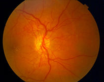 Diabetic retinopathy is a complication of diabetes that weakens the blood vessels that supply nourishment to the retina (the light-sensitive lining in the back of the eye where vision is focused). These weak vessels can leak, swell or develop thin branches, causing a loss of vision. Changes to your vision may not be noticeable at first. But in its advanced stages, the disease can cause blurred or cloudy vision, floaters and blind spots and, eventually, blindness. This damage is irreversible. Diabetic retinopathy is the most common diabetic eye complication and a leading cause of blindness in American adults. Macular edema, which is leaking fluid that causes blurred vision, often occurs with diabetic retinopathy.
Fortunately, diabetic retinopathy is preventable. People with diabetes are most susceptible to developing it, but your risk is reduced if you follow your prescribed diet and medications, exercise regularly, control your blood pressure, and avoid alcohol and cigarettes. Regular eye exams are an integral part of making sure your eyes are healthy. Diabetic retinopathy can be detected through a visual acuity test, a dilated eye exam or tonometry.
Although damage caused by diabetic retinopathy cannot be corrected, patients diagnosed with the condition can be treated to slow its progression and prevent further vision loss. Treatment modalities include laser and surgical procedures.
Flashes and Floaters
Flashes and floaters are symptoms of the eye that commonly occur as a result of age-related changes to the vitreous gel. When we are born, the vitreous is firmly attached to the retina and is a thick, firm substance without much movement. But as we age, the vitreous becomes thinner and more watery, and tissue debris that was once secure in the firm gel can now move around inside the eye, casting shadows on the retina.
Flashes in vision occur as a result of pressure on the retina in the back of the eye, and causes patients to see flashing lights or lightning streaks. Floaters occur when fibers move across the vitreous and into your field of vision, causing patients to see specks, strands, webs or other shapes as the fibers cast shadows on the retina. These spots are most visible when looking at a plain, light background.
Although flashes and floaters are common, especially as we age, it is important to see your doctor if you experience them, as they may indicate a retinal tear or hole. Your doctor can distinguish between harmless flashes and floaters, and those that may require treatment for an underlying condition. Most flashes and floaters will become less noticeable with time as patients adjust their vision. Although these floaters are harmless, it is important to continue to receive regular eye exams to ensure that any permanent changes to your vision do not occur.
Macular Degeneration
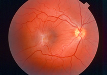
 The macula is a part of the retina in the back of the eye that ensures that our central vision is clear and sharp. Age-related macular degeneration (AMD) occurs when the arteries that nourish the retina harden. Deprived of nutrients, the retinal tissues begin to weaken and die, causing vision loss. Patients may experience anything from a blurry, gray or distorted area to a blind spot in the center of vision.
AMD is the number-one cause of vision loss in the U.S. Macular degeneration doesn’t cause total blindness because it doesn’t affect the peripheral vision. Possible risk factors include genetics, age, diet, smoking and sunlight exposure.Regular eye exams are highly recommended to detect macular degeneration early and prevent permanent vision loss.
Symptoms of macular degeneration include:
- A gradual loss of ability to see objects clearly
- A gradual loss of color vision
- Distorted or blurry vision
- A dark or empty area appearing in the center of vision
There are two kinds of AMD: wet (neovascular / exudative) and dry (non-neovascular). About 10-15% of people with AMD have the wet form. “Neovascular” means “new vessels.” Accordingly, wet AMD occurs when new blood vessels grow into the retina as the eye attempts to compensate for the blocked arteries.
These new vessels are very fragile, and often leak blood and fluid between the layers of the retina. Not only does this leakage distort vision, but when the blood dries, scar tissue forms on the retina as well. This creates a dark spot in the patient’s vision.
Dry AMD is much more common than wet AMD. Patients with this type of macular degeneration do not experience new vessel growth. Instead, symptoms include thinning of the retina, loss of retinal pigment and the formation of small, round particles inside the retina called drusen.
Vision loss with dry AMD is slower & often less severe than with wet AMD. Recent developments in ophthalmology allow doctors to treat many patients with early-stage AMD with the help of lasers and medication.
Amsler Grid
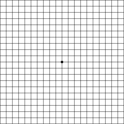
The Amsler Grid chart should be used to check the central part of your visual field, the area that can be damaged by Macular Degeneration.
Instructions for Use:
- View the chart (with your reading glasses on) at normal reading distance.
- Completely cover one eye and look at the central dot on the chart.
- Notice if there are any areas within the grid that appear gray (or black), or if there are any areas where the straight lines appear bent, crooked, or missing.
- Check the chart regularly (daily or weekly).
- Call your doctor if new changes occur.
Macular Pucker (Epiretinal Membrane)
 An epiretinal membrane, also known as a macular pucker, is a thin layer of scar tissue that forms over the macula, the area of the retina that gives us clear central vision. An epiretinal membrane often develops with age, as the vitreous gel that makes up most of the eye’s volume thins and pulls away from the retina. The damage caused to the retina leads to the formation of scar tissue on the retina. When the scar tissue contracts, the retina wrinkles, or puckers, causing blurry or distorted central vision.
Patients with an epiretinal membrane may experience difficulty seeing fine details and reading small print, and may also see straight lines appearing as wavy. There may also be a gray area or blind spot in the center of your vision. Although the cause of the conditions is similar, an epiretinal membrane is different from a macular hole. A macular hole is usually a much more serious condition that can progressively worsen.
Most cases of epiretinal membranes do not progress and do not require treatment. The symptoms of distortion and blurriness are usually mild and patients are able to adjust to the vision changes without much impact on their daily lives. Noninvasive treatments such as eye drops or medications will not improve vision that is distorted from an epiretinal membrane. If vision distortion is severe enough, a vitrectomy may be performed to repair this condition.
Macular Hole
Coming soon
Retinal Tear & Detachment
coming Soon